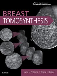
Providing unparalleled coverage, Breast Tomosynthesis explains how this new modality can lead to enhanced interpretation and better patient outcomes. It's an indispensable guide for those looking to keep abreast of the latest developments with correl ...view more
Providing unparalleled coverage, Breast Tomosynthesis explains how this new modality can lead to enhanced interpretation and better patient outcomes. It's an indispensable guide for those looking to keep abreast of the latest developments with correlative findings, practical interpretation tips, physics, and information on how tomosynthesis differs from conventional 2D FFDM mammography. Over 900 high-quality images offer visual guidance to effectively reading and interpreting this key imaging modality, while a separate section of Video Case Studies includes mammography images and over 90 tomosynthesis videos that allow users to see side-by-side findings and make comparisons using both modalities for the same patient case.
Providing unparalleled coverage, Breast Tomosynthesis explains how this new modality can lead to enhanced interpretation and better patient outcomes. It's an indispensable guide for those looking to keep abreast of the latest developments with correlative findings, practical interpretation tips, physics, and information on how tomosynthesis differs from conventional 2D FFDM mammography. Over 900 high-quality images offer visual guidance to effectively reading and interpreting this key imaging modality, while a separate section of VideoCase Studies includes mammography images and over 90 tomosynthesis videos that allow users to see side-by-side findings and make comparisons using both modalities for the same patient case.
Key Features
Includes over 900 high-quality tomosynthesis and mammographyimages representing the spectrum of breast imaging.
Features the latest Breast Imaging Reporting and Data System (or BI-RADS) standardsupdated in February 2014.
Highlights practical tips to interpreting this new modality and how it differs from 2D mammography.
Details how integration of tomosynthesis drastically changes lesion work-up and overall workflow in the department.
"Tomo Tips"boxes offer tips and pitfalls for expert clinical guidance.
A separate section of VideoCase Studies includes mammography images and over 90 tomosynthesis videos referenced online, allowing users to see side-by-side findings, including normal and benign and malignant lesions, and make comparisons using both modalities for the same patient case.
Expert Consult eBook version included with purchase. This enhanced eBook experience allows you to search all of the text, figures, images, videos, and references from the book on a variety of devices.
Author Information
By Liane E Philpotts, MD, FACR, Professor. Chief of Breast Imaging, Department of Radiology and Biomedical Imaging, Yale University School of Medicine, New Haven, Connecticut and Regina J Hooley, MD, Associate Professor, Vice Chair for Clinical Affairs, Department of Radiology and Biomedical Imaging, Yale University School of Medicine, New Haven, Connecticut
Bring it with you - The Bookshelf app allows you to access books on your laptop, tablet or mobile, so your ebooks go where ever you are - online or offline.
Take notes - Highlight, bookmark and take notes and highlights automatically stay in sync no matter where you make them.
Listen to eBooks - When you need to go screenless, the Text-to-speech tool will read your book aloud.
Powerful search - The searching capabilities allow you to search keywords through all your eBooks, the entire Bookshelf Library and well as on Wikipedia.
Ecological - Manage your environmental impact with paperless books.
How to read your VitalSource eBooks
Purchase your VitalSource eBook on the Elsevier Health site
https://www.uk.elsevierhealth.com/breast-tomosynthesis-9780323377447.html118723Breast Tomosynthesishttps://www.uk.elsevierhealth.com/media/catalog/product/9/7/9780323377447_8.jpg70.4993.99GBPInStock/Medicine and Surgery/Radiology/eBooks10029525504017453100005145120<p>Providing unparalleled coverage, <b><i>Breast Tomosynthesis</b></i> explains how this new modality can lead to enhanced interpretation and better patient outcomes. It's an <b>indispensable guide</b> for those looking to keep abreast of the<b> latest developments</b> with correlative findings, practical interpretation tips, physics, and information on how tomosynthesis differs from conventional 2D FFDM mammography. <b>Over 900 high-quality images</b> offer visual guidance to effectively reading and interpreting this key imaging modality, while a separate section of <b>Video</b> <b>Case Studies</b> includes mammography images and <b>over 90 tomosynthesis videos</b> that allow users to see side-by-side findings and make comparisons using both modalities for the same patient case.</p> <p>Providing unparalleled coverage, <b><i>Breast Tomosynthesis</b></i> explains how this new modality can lead to enhanced interpretation and better patient outcomes. It's an <b>indispensable guide</b> for those looking to keep abreast of the<b> latest developments</b> with correlative findings, practical interpretation tips, physics, and information on how tomosynthesis differs from conventional 2D FFDM mammography. <b>Over 900 high-quality images</b> offer visual guidance to effectively reading and interpreting this key imaging modality, while a separate section of <b>Video</b> <b>Case Studies</b> includes mammography images and <b>over 90 tomosynthesis videos</b> that allow users to see side-by-side findings and make comparisons using both modalities for the same patient case.</p>00add-to-cart97803233774472016ProfessionalBy Liane E Philpotts, MD, FACR and Regina J Hooley, MD20181E-BookOtherApprox. 750 illustrations (100 in full color)Elsevier35023 Dec 2016IN STOCKBy <STRONG>Liane E Philpotts</STRONG>, MD, FACR, Professor. Chief of Breast Imaging, Department of Radiology and Biomedical Imaging, Yale University School of Medicine, New Haven, Connecticut and <STRONG>Regina J Hooley</STRONG>, MD, Associate Professor, Vice Chair for Clinical Affairs, Department of Radiology and Biomedical Imaging, Yale University School of Medicine, New Haven, ConnecticuteBooksNoNoNoNoPlease SelectPlease SelectPlease Select